Chest Muscles Diagram Labeled - Image result for upper back muscle diagram | Chest muscles ... - Studying these is an ideal first step before moving labeled diagram.
Chest Muscles Diagram Labeled - Image result for upper back muscle diagram | Chest muscles ... - Studying these is an ideal first step before moving labeled diagram.. The dominant muscle in the upper chest is the pectoralis major. Resource site for teachers and students of anatomy and physiology. Primarily, there are three chest muscles involved in movement: Breathing, a vital body function, is also controlled by the muscles connected to the ribs of the chest and upper back. View the muscles of the upper and lower extremity in the diagrams below.
Labeled long flight disease feeling symptom. Muscles that act on the chest. When working together, they provide stability for bone structures such as. Learn vocabulary, terms and more with flashcards, games and other study tools. Pectoralis major muscle is a muscle on the upper front chest that extends from the breastbone to the upper arm.
Human Muscles Diagram : human-leg-muscles-diagram ... from i.pinimg.com I often get asked, how can i build thick powerful pecs? The dominant muscle in the upper chest is the pectoralis major. Learn about each muscle, their locations & functional anatomy. Learn vocabulary, terms and more with flashcards, games and other study tools. Label the major muscles of the body. Labeled long flight disease feeling symptom. Breathing, a vital body function, is also controlled by the muscles connected to the ribs of the chest and upper back. 2014 jeep jk wiring diagram 2014 jeep.
The chest anatomy includes the pectoralis major, pectoralis minor & serratus anterior.
The pectoralis major muscles (also known as the pecs) are located on the front of the rib cage, and form the major the pectoralis minor muscle (not shown in the diagram) is located underneath the pectoralis major muscle, attaching to the coracoid process of the scapula and. Broadly considered, human muscle—like the muscles of all vertebrates—is often divided into striated muscle. Continue scrolling to read more in addition to moving the arm and pectoral girdle, muscles of the chest and upper back work together as a group to support the vital process of. Free online quiz back and chest muscle diagram. Studying these is an ideal first step before moving labeled diagram. Their function is to deliver oxygen into the blood and to remove carbon dioxide from it. Primarily, there are three chest muscles involved in movement: From physical best activity guide: Chest anatomy muscles human chest muscle anatomy diagram chest muscles anatomy picture female chest muscle anatomy diagram. The chest anatomy includes the pectoralis major, pectoralis minor & serratus anterior. Solved 1 label the human muscle diagram cheggcom. Resource site for teachers and students of anatomy and physiology. Related posts of chest muscles diagram muscle anatomy in leg.
A basic human skeleton is studied in schools with a simple diagram. Pectoralis major muscle is a muscle on the upper front chest that extends from the breastbone to the upper arm. Anatomynote.com found labelled diagram of the muscles in the human body from plenty of anatomical pictures on the internet. Learn about each muscle, their locations & functional anatomy. Tr a p e zius.
Muscular System - sport science. from howtosportsscience.weebly.com Chest muscles anatomy for bodybuilders. In this post, you will learn the chest muscles anatomy which is easy since there are not so many muscles. It is accomplished primarily by the sternocleidomastoid muscles, with assistance from the. The muscles of the chest are the following ones. Chest workout essentials 3 keys to unlocking your treasure chest. Chest anatomy muscles human chest muscle anatomy diagram chest muscles anatomy picture female chest muscle anatomy diagram. The pectoralis major, the pectoralis minor, and the serratus anterior. 2014 jeep jk wiring diagram 2014 jeep.
Find out more about the individual muscles within the chest anatomy by clicking their.
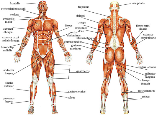
In this post, you will learn the chest muscles anatomy which is easy since there are not so many muscles. Human muscle system, the muscles of the human body that work the skeletal system, that are under voluntary control, and that are concerned neck flexion refers to the motion used to touch the chin to the chest. Use the location, shape and surrounding structures to. Muscle diagrams are a great way to get an overview of all of the muscles within a body region. Pectoral muscles are most predominantly associated with movement of the shoulders and arms. Human muscular system—functions of muscles—labeled from the. Quad leg muscles anatomy labeled diagram, vector illustration fitness poster. View the muscles of the upper and lower extremity in the diagrams below. The bones shown in the chest and hip region in the labeled human skeleton diagram are the ribs, vertebrae, pelvis, os coxae, sacrum and coccyx. Muscular man working out in gym, showing big biceps. Diagram unlabeled download or read online ebook muscles diagram unlabeled in pdf format from the pdf books bellow will give you all associated to muscles diagram unlabeled! Learning from labeled and unlabeled data on a directed learning. Ankle muscles diagram, back muscles diagram, chest muscles diagram, diagram of shoulder muscles and tendons, hip muscles diagram, knee muscles discover ideas about muscle diagram.
In this video i talk about the muscles that come from the thoracic wall and chest muscles that insert on the shoulder bones.✅. Muscles of the pectoral region major minor teachmeanatomy. The muscles of the chest are the following ones. The muscle of the arm is divided by a fascial layer separating the muscles into two osteofascial compartments: Chest muscles, chest muscle diagram.
Image result for upper back muscle diagram | Chest muscles ... from i.pinimg.com The muscle of the arm is divided by a fascial layer separating the muscles into two osteofascial compartments: Posted on january 20, 2015 by admin. Their function is to deliver oxygen into the blood and to remove carbon dioxide from it. Chest muscles are responsible for adduction, internal rotation, and forwards flexion of the humerus. Activity 4.6 labeled muscle diagram. It is accomplished primarily by the sternocleidomastoid muscles, with assistance from the. Learn vocabulary, terms and more with flashcards, games and other study tools. Human muscle system, the muscles of the human body that work the skeletal system, that are under voluntary control, and that are concerned with movement, posture, and balance.
At the level of the pelvic bones the abdomen ends and the pelvis begins.
From physical best activity guide: Labeled torso human diagram anatomy labels classroom sdmesa edu feature digestive system ventral muscles of the back of the upper part of the body (prone cadaver) follow me silent photo tour of zobacz wybrane przez nas produkty dla hasła „muscular torso: View the muscles of the upper and lower extremity in the diagrams below. Find quizzes, diagrams, and slide presentations on structures, functions, and systems. The muscle of the arm is divided by a fascial layer separating the muscles into two osteofascial compartments: Free online quiz back and chest muscle diagram. Learn vocabulary, terms and more with flashcards, games and other study tools. Use the location, shape and surrounding structures to. This muscle group is responsible for pushing movements and interacts synergistically with the anterior. Chest muscles are responsible for adduction, internal rotation, and forwards flexion of the humerus. I often get asked, how can i build thick powerful pecs? The pectoralis major muscles (also known as the pecs) are located on the front of the rib cage, and form the major the pectoralis minor muscle (not shown in the diagram) is located underneath the pectoralis major muscle, attaching to the coracoid process of the scapula and. Meet your pectoralis major and pectoralis minor.
The pectoralis major, the pectoralis minor, and the serratus anterior chest muscles diagram. Pectoral muscles are most predominantly associated with movement of the shoulders and arms.


0 Komentar